Spiga

6 steps to longer life,healthy heart

6 Steps to Longer Life, Healthy Heart

These Strategies Could Prevent More Than 27 Million Heart Attacks, Experts Estimate

By Miranda Hitti
WebMD Medical News


Reviewed By Louise Chang, MD

July 7, 2008 — Living longer, with a healthier heart, boils down to a few steps, and if everyone got on the bandwagon, it could prevent more than 27 million heart attacks and about 10 million strokes over the next 30 years.

That news comes from a study backed by the American Heart Association, American Diabetes Association, and American Cancer Society.

Here are those steps:

  • Quit smoking
  • Get your BMI (body mass index) out of the obese range
  • Get your LDL ("bad") cholesterol under control
  • Get your blood pressure into the normal range
  • Get your blood sugar under control, especially if you have diabetes
  • Take aspirin if your doctor tells you to because of heart disease risk

If you're like 78% of U.S. adults aged 20-80, you fall short of at least one of those goals.

If everyone who's got room for improvement made those improvements and met all of the benchmarks, they would live 1.3 years longer, on average, cutting heart attacks by 63% and stroke by 31% over the next 30 years, the researchers estimate.

But let's say everyone makes some progress but doesn't quite meet all of the goals. That would still make a real dent in cardiovascular disease, the No. 1 killer of U.S. men and women, according to the study.

The study, published in the advance online edition of Circulation, is based on national health studies conducted from 1998 to 2004.

Are medications the answer? If everyone relied on prescription drugs, it could be a hefty tab. But lifestyle changes — such as a healthier diet and a more active lifestyle — are a big part of heart health. The researchers didn't crunch the numbers to see how many people could meet most of the goals through lifestyle change alone.


Frequent sex protects against erectile dysfunction

Erections: Use 'Em or Lose 'Em

Frequent Sex Protects Against Erectile Dysfunction

By Daniel J. DeNoon
WebMD Medical News

Reviewed By Louise Chang, MD


July 3, 2008 — Men who don't use their erections lose them, Finnish researchers find.

Aging men who have sex at least once a week have only half the risk of developing erectile dysfunction as do men who have sex less often.

But once-a-weekers shouldn't gloat. More sex means even less ED risk. Men who have sex at least three times a week are only one-fourth as likely to get erectile dysfunction as are men who have less-than-weekly sex.

"Regular sexual activity preserves potency in a similar fashion as physical exercise maintains functional capacity," conclude Juha Koskimaki, MD, PhD, and colleagues at the University of Tampere, Finland.

The findings come from questionnaires mailed to Finnish men aged 55 to 75. Only the 989 men who did not have erectile dysfunction at the beginning of the study — and who returned a second questionnaire five years later — were included.

Men with erectile dysfunction obviously have sex less often than do more potent men. But by including only men who did not have erectile dysfunction to start with, Koskimaki and colleagues believe their study strongly suggests that sexual intercourse lowers the risk of ED.

The average man in the study was 59 years old. Four out of five of the men were married or cohabitating. More than half of them were overweight, and nearly half had at least one chronic medical condition.

For such men, Koskimaki and colleagues find, sex less than once a week significantly increases the risk of erectile dysfunction. And compared to sex once a week, sex at least three times a week significantly decreases risk of erectile dysfunction.

Interestingly, the study found that men who have less than one morning erection per week are 2.5 times more likely to get erectile dysfunction as are men who have two or three morning erections per week. But having a morning erection every day did not lower a man's risk of erectile dysfunction.

One major limitation of the study, Koskimaki and colleagues note, is that they did not ask the men about masturbation, which might conceivably have the same salubrious effect on erectile dysfunction as intercourse. So as far as the researchers can tell, the study findings apply only to sex with another person.

"Doctors should support patients' sexual activity," they conclude.

Koskimaki and colleagues report their findings in the July 2008 issue of The American Journal of Medicine.


Dedah aurat untuk perubatan

Soalan: Isteri saya mengalami masalah kegemukan. Saya berhasrat ingin menghantar dia ke pusat rawatan menguruskan badan. Masalahnya, kebanyakan pusat-pusat menguruskan badan yang baik dikendalikan oleh wanita-wanita berbangsa Cina (bukan Islam). Mungkin timbul di sana masalah aurat terdedah semasa rawatan. Apakah hukum mendedahkan aurat kepada bukan muslim untuk tujuan rawatan seperti di atas.?


Jawapan:
Pertama, saudara perlu tahu had aurat seorang lelaki dan wanita. Mereka tidak boleh membuka aurat atau membenarkan ia disentuh kecuali atas sebab yang dibenarkan dalam syarak. Aurat paling berat ialah qubul dan dubur. Manakala aurat paling ringan seperti kedua-dua belah peha lelaki terdedah sesama mereka.
Kedua, menurut kaedah usul fikah diharuskan melakukan perkara yang diharamkan dalam syarak sekiranya boleh mendatangkan mudarat. Oleh itu, diharuskan membuka aurat untuk tujuan menguruskan badan kerana kegemukan boleh mendatangkan pelbagai penyakit. Ulama tidak berselisih pendapat mengharuskan doktor lelaki melihat aurat pesakit wanita. Begitu juga sebaliknya dengan syarat mereka hendaklah amanah dan menutup keaiban pesakit itu.
Cadangan saya agar saudara mendahulukan doktor perempuan atau pusat perubatan Islam sebelum menemui doktor lelaki atau pusat perubatan bukan Islam. Setelah berusaha, tidak mengapa menggunakan perkhidmatan mereka. Wallahu a’lam

Sumber : Soal Jawab Agama ALAF 21, http://agama.zonalaf.com.my


Promising Cancer Treatment Ready for Human Trial


Promising Cancer Treatment Ready for Human Trial

MONDAY, June 30 (HealthDay News) — A clinical trial will examine whether a new cancer treatment is as effective in humans as it's proven to be in mice, say researchers at Wake Forest University Baptist Medical Center in Winston-Salem, N.C.

The treatment involves transfusing white blood cells called granulocytes from healthy young donors — whose immune systems produce cells with high levels of cancer-fighting activity — into patients with advanced cancer.

A similar treatment using white blood cells from cancer-resistant mice cured 100 percent of lab mice with advanced cancer.

"In mice, we've been able to eradicate even highly aggressive forms of malignancy with extremely large tumors. Hopefully, we will see the same results in humans. Our laboratory studies indicate that this cancer-fighting ability is even stronger in healthy humans," lead researcher Zheng Cui, associate professor of pathology, said in a prepare statement.

The researchers will select 100 healthy donors, age 50 or younger, who have white blood cells with high cancer-killing activities. The recipients will included 22 patients with solid tumors that aren't responding to conventional therapy.

"If the study is effective, it would be another arrow in the quiver of treatments aimed at cancer," co-researcher Dr. Mark Willingham, a professor of pathology, said in a prepared statement. "It is based on 10 years of work since the cancer-resistant mouse was first discovered."

This phase II study is designed to determine whether cancer patients can tolerate a sufficient amount of transfused granulocytes for treatment. After three months, the patients will be evaluated to determine whether the treatment provided clear clinical benefits.

Details of the study were presented June 28 at the Understanding Aging conference in Los Angeles. If this trial proves successful, the researchers will then look at whether this treatment is best suited for treating certain types of cancer.

— Robert Preidt

Deep Vein Thrombosis : life threat


Signs and symptoms of DVT:
  • pain in the leg
  • tenderness in the calf
  • swelling of the leg
  • increased warmth of the leg
  • erythema ( redness) of the leg
  • bluish skin discoloration

Watermelon : a natural viagra?


Watermelon: A Natural Viagra?

Researcher Says Popular Summer Fruit May Have Viagra-Like Effect on Blood Vessels

By Kathleen Doheny
WebMD Health News Reviewed by Brunilda Nazario, MD


July 1, 2008 -- Men hoping for some fireworks in their love life this Fourth of July may want to skip the burgers and beer at the barbecue and eat plenty of watermelon.

Watermelon may be a natural Viagra, says a researcher. That's because the popular summer fruit is richer than experts believed in an amino acid called citrulline, which relaxes and dilates blood vessels much like Viagra and other drugs meant to treat erectile dysfunction (ED).

"We have known that watermelon has citrulline," says Bhimu Patil, PHD, director of the Fruit and Vegetable Improvement Center at Texas A&M University, College Station. Until recently, he tells WebMD, scientists thought most of the citrulline was in the watermelon rind. "Watermelon has more citrulline in the edible part than previously believed," he says.

How could watermelon be a natural Viagra? The amino acid citrulline is converted into the amino acid arginine, Patil says. "This is a precursor for nitric oxide, and the nitric oxide will help in blood vessel dilation."

So, the burning question: How much watermelon does it take?

"That is a good question," Patil says. Unfortunately, "I don't have an answer for that."

He does know that a typical 4-ounce serving of watermelon (about 10 watermelon balls) has about 150 milligrams of citrulline. But he can't say how much citrulline is needed to have Viagra-like effects.

He's hopeful that someone will pick up on his research and study the fruit's effect on penile erections.

Watermelon's Viagra-Like Effects
On hearing about the Texas finding, Irwin Goldstein, MD, editor-in-chief of The Journal of Sexual Medicine, was underwhelmed. Suggesting a man feast on watermelon to boost performance, he says, "would be the equivalent of someone dropping a beer bottle in Minneapolis, where the Mississippi River starts, and hoping to see it make an impact on someone in New Orleans."

"To say that watermelon is Viagra-like is sort of fun," says Goldstein. "But to even vaguely hope that eating watermelon will alleviate ED is misleading."

"The vast majority of Americans produce enough arginine," adds Goldstein, medical director of Alvarado Hospital Medical Center, San Diego, and clinical professor of surgery, University of California San Diego School of Medicine. "Men with ED are not deficient in arginine."

Though arginine is required to make nitric oxide, and nitric oxide is required to dilate blood vessels and have an erection, "that doesn't mean eating something that is rich in citrulline will make enough arginine that it will lead to better penile erections," Goldstein says.

Goldstein has served as a consultant for many companies that make ED drugs.

Calling watermelon a natural Viagra is "clearly premature," says Roger Clemens, DrPH, adjunct professor of pharmacology and pharmaceutical sciences, University of Southern California, Los Angeles, and a spokesman for the Institute of Food Technologists.

Clemens studied the amino acid arginine himself, researching a supplement to improve vascular flow for patients with hardening of the arteries or atherosclerosis. He has since abandoned that line of research, he says.

It can require a lot of watermelon to boost blood levels of arginine, he adds. In a study published in 2007 in Nutrition, he says, volunteers who drank three 8-ounce glasses of watermelon juice daily for three weeks boosted their arginine levels by 11%.

Watermelon is low in calories and provides potassium and the phytonutrients lycopene and beta-carotene, in addition to the citrulline.

Clemens' advice on watermelon and the Fourth of July? "Put salt on it and enjoy."

Just don't expect fireworks anywhere but in the sky.

Achalasia

Achalasia


Definition : Primary oesophageal motility disorder , characterized by failure of the lower oesophageal sphincter to relax and absence of oesophageal peristalsis

Symptoms:
  • dysphagia ( both liquid and solid)
  • heartburn
  • regurgitation
  • chest pain
  • weight loss
Investigation:

a) Imaging :
  • barium swallow: oesophagus appears dilated , contrast material passes slowly into the stomach as the LES opens intermittenly .Distal oesophagus is narrowed( resembling bird`s beak)

b) Prodecural
  • OGDS  : to rule out ca of gastroeophageal junction or fundus
c) other test
  • oesophageal manometry : to diagnose achalasia
  1. incomplete relaxation of LES in response to swallowing
  2. high resting LES pressure
  3. absent of oesophageal peristalsis
  • prolonged oesophageal pH monitoring : to rule out GERD
Treatment
  • reduce LES pressure : calcium channel blockers and nitrates
  • intrasphincteric injection of botulinum toxin : restore balance between excitatory and inhibitory neurotransmitters
  • pneumatic dilatation surgery : balloon inflated at the gastroesophageal junction to rupture the muscle fibers 

Green Tea Lowers Risk of Heart Disease

Green Tea Lowers Risk of Heart Disease
Drinking Green Tea Boosts the Health of Blood Vessels Within Minutes

By Kelli Miller Stacy
WebMD Medical News

Reviewed By Elizabeth Klodas, MD, FACC

July 2, 2008 — Drinking green tea rapidly improves the health of the delicate cells lining the blood vessels and helps lower one's risk of heart disease.

Researchers writing in the latest issue of the European Journal of Cardiovascular Prevention and Rehabilitation have found that people who drink green tea have better blood vessel function just 30 minutes later. Specifically, green tea improves the function of endothelial cells. Endothelial cell dysfunction plays a key role in the development of clogged arteries, a process called atherosclerosis.

The finding adds to a growing body of evidence that suggests that powerful antioxidants in green tea called flavonoids may protect the heart. Other flavonoid-rich foods include red grapes, red wine, and dark chocolate. The study authors say their results are the first to show that green tea offers a short-term improvement in the health of arteries. Black tea has previously been linked to short- and long-term improvements in endothelial function.

For the study, Nikolaos Alexopoulos and colleagues at the Athens Medical School in Greece randomly assigned 14 healthy volunteers approximately 30 years of age to a cup of green tea, a beverage containing the same amount of caffeine as green tea, or hot water on three separate occasions.

The researchers used a technique called flow-mediated dilation (FMD) to measure blood flow in each participant's arm at 30, 90, and 120 minutes after they drank their beverage. FMD is a noninvasive test that uses a blood pressure cuff and ultrasound to see how blood flows in the brachial artery when the arm is gently squeezed. The brachial artery runs from the shoulder to the elbow. The artery should get wider when blood flow in the area increases, but diseases such as atherosclerosis hamper this effect. FMD is an independent predictor of endothelial function and heart disease risk.

After drinking green tea, the subjects experienced significantly increased artery widening (dilation), with the highest increase noted at 30 minutes. The caffeinated beverage and hot water did not produce any significant changes in the same individuals.

SOURCES: News release, European Society of Cardiology. Alexopoulos, N. European Journal of Cardiovascular Prevention and Rehabilitation, June 2008: vol 15: pp 300-305.

Renal tubular acidosis


A 37-year-old man was referred for evaluation of distal renal tubular acidosis. Laboratory evaluation revealed a serum potassium level of 3.3 mmol per liter, a bicarbonate level of 16 mmol per liter, a calcium level of 9.3 mg per deciliter (2.3 mmol per liter), a phosphate level of 2.1 mg per deciliter (0.7 mmol per liter), a creatinine level of 3.0 mg per deciliter (265 µmol per liter), a parathyroid hormone level of 62 pg per milliliter, and an estimated glomerular filtration rate of 25 ml per minute per 1.73 m2 of body-surface area. He had been given a diagnosis of renal tubular acidosis at 9 years of age on the basis of metabolic acidosis with a high urinary pH and hypokalemia associated with nephrocalcinosis. At that time, there was evidence of bilateral nephrocalcinosis on plain abdominal radiography. The patient was treated with sodium bicarbonate and potassium supplementation and had normal growth but did not undergo medical follow-up or treatment between 15 and 37 years of age. The plain film of the abdomen obtained during the referral visit (see figure) revealed bilateral symmetric calcification of the renal parenchyma, sparing only the renal pelvis. This finding contrasts with those classically associated with type 1 distal renal tubular acidosis, in which nephrocalcinosis is present but is limited to the renal medulla. Three years after sodium bicarbonate and potassium supplementation was restarted, the patient's renal function has remained stable.

by:
Andres Serrano, M.D.
Daniel Batlle, M.D.
Northwestern University Feinberg School of Medicine
Chicago, IL 60611

Intestinal obstruction

Cardinal features of intestinal obstruction

  • vomiting
  • constipation
  • abdominal pain
  • abdominal distension


Coronary arteries and the tributaries

Respiratory cause of clubbing

Respiratory cause of clubbing ( BECA MAD)


  • Bronchiectasis
  • Empyema
  • Cancer ( lung ca)
  • Asbestosis
  • Mesothelioma
  • Abscess( lung abscess)
  • Drugs

alpha and beta adrenoreceptors


Alpha and Beta Receptors

  • epinephrine reacts with both alpha and beta adrenoreceptors
  • Alpha receptors stimulation cause vasoconstriction
  • Beta receptors stimulation cause vasodilation
  • alpha receptors are less sensitive to epinephrine but when activated,they override the vasodilation by beta receptors
  • high levels of epinephrine cause vasoconstriction
  • low levels of epinephrine will cause vasodilation

alpha 1 receptors
  • cause smooth muscle contraction which include blood vessels , skin, gastrointestinal system,renal artery and brain
  • induce glycogenolysis and gluconeogenesis in adipose tissue and liver cells
  • induce sodium reabsorption in the kidney
  • induce sweat glands secretion
alpha 2 receptors
  • inhibition of insulin release from pancreas
  • induce glucagon release from pancreas
  • induce contraction of gastrointestinal tract sphinter
beta 1 receptors
  • increase cardiac output : increase HR and increase contraction ( increase ejection fraction)
  • renin release from juxtaglomerular cells
  • lipolysis in adipose tissue
beta 2 receptors
  • smooth muscle relaxation eg: bronchodilator
  • dilate arteries to skeletal muscle
  • glycogenolysis and gluconeogenesis
  • increase renin secretion from kidney
  • inhibit histamine release from mast cells
beta 3 receptors
  • induce lipolysis

Pheochromocytoma


Pheochromocytoma 


Definition : Catecholamine secreting tumour derived from chromaffin cells of adrenal gland.

Pathophysiology : tumour secretes catecholamine  which includes epinephrine, norepinephrine and rarely dopamine
Effect :
  • Stimulation of alpha adrenergic receptors cause : elevated blood pressure, increased cardiac contractility, glycogenolysis, gluconeogenesis and intestinal relaxation
  • Stimulation of beta adrenergic receptors cause : increase heart rate and contractility
Symptoms:
  • Headache
  • palpitation
  • diaphoresis
  • severe hypertension
  • Constipation
  • anxiety
  • Flank pain 
  • epigastric pain
  • nausea
  • weight loss
  • tremor
Signs:
  • weight loss
  • neurofibroma
  • cafe au lait spots
  • pallor
  • tremor
  • fever hypertension
  • tachyarrhythmias
  • pulmonary odema
  • cardiomyopathy
  • ileus

Small bowel obstruction


Figure 1 : normal bowel

Figure 2 : Small bowel obstruction. multiple step ladder pattern of air-fluid level in the intestine

basic x ray

What to see from an x  ray film :


  • Check for name, age of patient
  • Check type of x ray : PA/AP/Supine/Lateral
  • Spine : central , any fracture?
  • Bones and ribs : any fracture
  • mediastinal contours
  • Trachea : should be central
  • Aortic arch , left pulmonary artery
  • Heart  : two thirds of heart lies on the left of the chest, one third on the right. The heart should take up no more than half on the thoracic cavity ( if yes : cardiomegaly ). 
  1. Left border of the heart is made up by the left atrium and left ventricle. 
  2. Right border is made up by the right atrium alone. Above he right heart border lies the edge of the superior vena cava
  • Pulmonary arteries and main bronchi : arise at the left and right hila . Bulky hilum maybe due to enlarged lymph nodes or tumours
  • Lungs : should be black ( if pneumothorax : sharp line of the edge of the lung)
  • Diaphragm : 
  1. check for surface of hemidraphragms curving downwards
  2. check for costophrenic angle : if blunted : effusion
  • Soft tissue : any injury


Scabies( Kudis Buta)

Scabies (Kudis Buta)


Organism : Sarcoptes scabei

Transmission : 
  • skin-to-skin contact, 
  • sharing of clothes, 
  • sharing of towels etc
( usually occurs in crowded areas and especially in boarding schools)


Symptoms:
  • pimple like irritations,burrows or rash of the skin ,especially the webbing between the fingers , the skin folds on the wrist, elbow or knee, penis , breast and or shoulder blades
  • intense itching 
  • sores on the body caused by scratching . It may get infected with bacteria
Investigations:

  • Skin scrapping : preferably from primary lesions
  • Burrow ink test
  • Topical tetracycline solution : examine under Wood`s light
Treatment: 

  • Scabicides : permethrin ; apply from chin to toes and under fingernails and toenails, rinse off in shower 12 hour later, repeat in 1 week
  • Scabicides : lindane : apply from chin to toes and under fingernails and toenails , rinse off in shower 10 hour later, repeat in 1 week.
  • all family members and close contacts must be evaluated and treated,even if they do not have symptoms
  • Patient to wash their clothes,bed lines and towels in hot water the day after treatment is initiated and again in 1 week.

Causes of crackles

Causes of crackles (BEABFO)

  • Early – Bronchitis, Emphysema, Asthma
  • Mid – Bronchiectasis (altered by coughing)
  • Late – Fibrosing alveolitis, pulmonary Oedema

Primary and Secondary survey

Primary Survey (ABCDE)

  • Airway
  • Breathing
  • Circulation and control of haemorrhage
  • Disability/deficit neurologically
  • Exposure

Secondary Survey (AMPLE)
  • Age/Allergies
  • Medication
  • Past Medical History
  • Last meal
  • Events/Exposure

Causes of acute pancreatitis

Causes of acute pancreatitis

  • G all stone
  • E thanol
  • T rauma
  • S teroids
  • M umps
  • A utoimmune (PAN
  • S corpion bite
  • H yperlipidaemia
  • H yperparathyroidism
  • H ypothermia
  • E RCP
  • D rugs

Varicose vein


<
Varicose vein : Elongated,tortuous dilated superficial veins

Risk factors of VV:

  • Female
  • age > 50 years old
  • Multiparrous
  • pregnancy
  • oral contraceptive pills
  • congenital
  • prolong standing

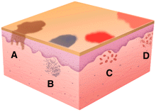
Macule& Patch


macule : A small, flat, distinct colored area of skin that is <1 cm in diameter
Patch : a flat,distinct colored area of skin that is > 1cm in diameter

Blood supply of the brain

The Carotid Circulation Supplies:
• optic nerves and retina
• cortex and deep white matter of the frontal and parietal lobes, and lateral aspects ofthe temporal and occipital lobes
• all of the corpus callosum except its posterior regions
• most of the basal ganglia and internal capsule

The right common carotid artery originates from the bifurcation of the brachiocephalic trunk,while the left common carotid originates directly from the aortic arch. Each common carotid thenbranches to form the internal and external carotid vessels. After the internal carotid ascendsthrough the neck, traverses the temporal bone, and passes through the cavernous sinus it finallyreaches the subarachnoid space at the base of the brain.

As the internal carotid leaves the cavernous sinus it gives rise to its first intracranial branch, theophthalmic artery, which travels along the optic nerve into the orbit. There its branches supplythe retina and other structures of the eyeball itself, as well as other structures in and around theorbit. The internal carotid continues in a superior direction and usually gives off two additionalbranches: the posterior communicating and anterior choroidal arteries.

The posterior communicating arteries usually link the internal carotid to the posterior cerebralartery, and may be large or threadlike. However, in a number of individuals one or both of theposterior cerebral arteries retain their embryological state as direct branches of the internalcarotid artery itself. The anterior choroidal artery also varies a great deal in size and importancein different individuals, and may branch from the middle cerebral artery rather than the internalcarotid. For this reason, we will discuss it with the middle cerebral artery. Finally, the internalcarotid divides to form the anterior and middle cerebral arteries.

Anterior Cerebral Artery

The anterior cerebral artery (ACA) arises from the internal carotid at nearly a right angle. Itsends deep penetrating branches to supply the most anterior portions of the basal ganglia. It thensweeps forward into the interhemispheric fissure, and then runs up and over the genu of thecorpus callosum before turning backwards along the corpus callosum. As it runs backwards itforms one branch that stays immediately adjacent to the corpus callosum while a second branchruns in the cingulate sulcus (just superior to the cingulate gyrus

ACA supplies the medial and superior parts of the frontal lobe, and of the anterior parietal lobe.

These regions include the following key functional areas:
• septal area
• primary motor cortex for the leg and foot areas, and the urinary bladder
• additional motor planning areas in the medial frontal lobe, anterior to the precentral gryus
• primary somatosensory cortex for the leg and foot

ACA also supplies most of the corpus callosum except its posterior part. These callosal fibersenable the language-dominant hemisphere to find out what the other hemisphere is doing, and todirect its activities

The short anterior communicating artery joins the two anterior cerebral arteries. This vessel mayallow collateral flow into the opposite hemisphere if the carotid artery is occluded on either side

Middle Cerebral Artery
The middle cerebral artery (MCA) has a large diameter and branches at an acute angle fromthe internal carotid. The MCA passes laterally just underneath the frontal lobe, ultimately takingup a position between the temporal and frontal lobes in the Sylvian fissure. The initial part of theMCA is a single vessel called the stem or M1 segment. As it passes laterally, the stem gives off aseries of 6-12 long, small diameter penetrating vessels that travel directly upward to supply thebasal ganglia and much of the internal capsule. These are called the lenticulostriate arteries.

The lenticulostriate vessels are small diameter arteries that originate as right angle branchesfrom the MCA stem (a large diameter vessel with a brisk, high pressure blood flow). These smallarteries are particularly susceptible to damage from hypertension. They may either rupture(producing an intracerebral hemorrhage that is initially centered in the region they supply) orbecome occluded (producing a lacunar infarct in the tissue they supply). The lenticulostriatearteries are ‘end arteries’ and regions that they supply do not have significant collateral bloodsupply. Therefore occlusion of these vessels leads to stereotyped stroke syndromes.

In the case of the lenticulostriate vessels, hemorrhage may remain localized to the putamen (andcaudate), may involve neighboring structures like the internal capsule and other more distantwhite matter of the hemisphere, or may even rupture into the ventricular system. Lacunarinfarcts may have serious functional consequences if they involve motor or sensory fibers in theinternal capsule but may be ‘silent’ if they involve other small regions of white matter or thebasal ganglia.Once in the Sylvian fissure itself, the MCA stem divides into two or, in a smaller number ofcases, three main cortical branches that supply almost the entire lateral surface of the brain aswell as the insula. Large emboli carried up the carotid tend to be swept into MCA, and are proneto getting stuck at this branch point.

The superior (upper or suprasylvian) MCA branch gives rise to several arteries that supply muchof the lateral and inferior frontal lobe and the anterior lateral parts of the parietal lobe. Theinferior (lower or infrasylvian) MCA branch gives rise to arteries that supply the lateral temporallobe including its anterior tip and the amygdala, posterior parietal and much of the lateraloccipital lobe. Emboli can also lodge in one of these two major cortical branches, as well as inthe smaller arteries which each of them will subsequently form..

There is a tendency for atheromatous plaques to form at branchings and curves of thecerebral arteries. Thus in the carotid circulation the most frequent sites are in the internalcarotid artery at its origin from the common carotid, in the stem MCA or its bifurcation intosuperior and inferior divisions, or in the ACA as it curves backwards over the corpuscallosum.

Superior branches of MCA participate in supplying the following key functional areas:
• Primary motor cortex for face and arm, and axons originating in the leg as well asface and arm areas that are headed for the internal capsule as part of the corticobularor corticospinal tracts
• Broca’s area and other related gray and white matter important for languageexpression -- in the language-dominant (usually L) hemisphere
• Frontal eye fields (important for ‘looking at’ eye movements to the opposite side)
• Primary somatosensory cortex for face and arm • Parts of lateral frontal and parietal lobes important for 3-D visuospatial perceptionsof one’s own body and of the outside world, and for the ability to interpret andexpress emotions -- in the nondominant (usually R) hemisphere

Inferior branches of MCA participate in supplying the following key functional areas:
• Wernicke’s and other related areas important for language comprehension in thelanguage- dominant (usually L) hemisphere • Parts of the posterior parietal lobe important for 3-D visuospatial perceptionsperceptions of one’s own body and of the outside world, and for the ability tointerpret emotions -- in the nondominant (usually R) hemisphere
• Optic radiations, particularly fibers that represent information from the contralateralsuperior quadrants and loop forward into the temporal lobe (they are located anteriorand lateral to the temporal horn of the lateral ventricle) as they travel from the lateralgeniculate body to the striate cortex, located in the occipital lobe